Physical Description

|
|
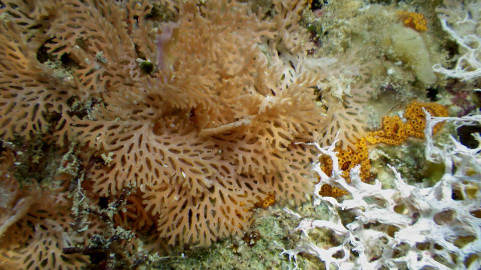
Photo: Bridget Bradshaw, Heron Island Reef, 2013. |
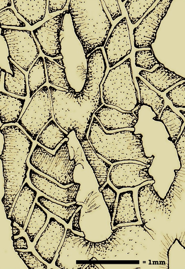
R. graeffei belongs to the family Phidoloporidae, which may be distinguished from other bryozoans most obviously by their lace-like appearance, though some genera do not form the characteristic networks. Additionally, phidoloporids lack zooids on the basal side of their skeleton (see Fig. 1), leaving the apical side peppered with opercula of the individual zooids. The delicate fenestrate structure becomes more dimensionally complex with size, the larger colonies forming wavy rosettes with multiple layers of fan-like branches. The colony attaches to the substrate with a kenozooid (a zooid specialized as a holdfast) and radiates outwards (Ryland 1970). In larger colonies, the proximal (closest to anchor point) end of the branches tends to be colorless while the distal edge of the colony appears pinkish-orange. This seems to be a result of a higher density of embryos (see Embryo density, REPRODUCTION) farther from the anchor.

|
| Figure 1. R. graeffei, basal side. |
Based on personal observation, colony size appears to be highly variable and is likely a function of age (Ryland 1970), with some small colonies only a few centimeters in diameter with one layer of branches while older colonies reach 7-8 cm in diameter with more elaborately layered branches. They tend to be brittle and the apical tips break easily. Lophophores will retract if jostled or if left for a period of time in still water, but when protruding give the colony a fuzzy look.
The carbonate structure of the colony is diagnostic within cheliostome bryozoan species, and is best seen using scanning electron microscopy (SEM). In a series of SEM photos taken by Kevin Tilbrook (2006), he describes the features of the frontal avicularia (specialized defense zooid), orifice shape and number of peristomial spines unique to R. graeffei.

|
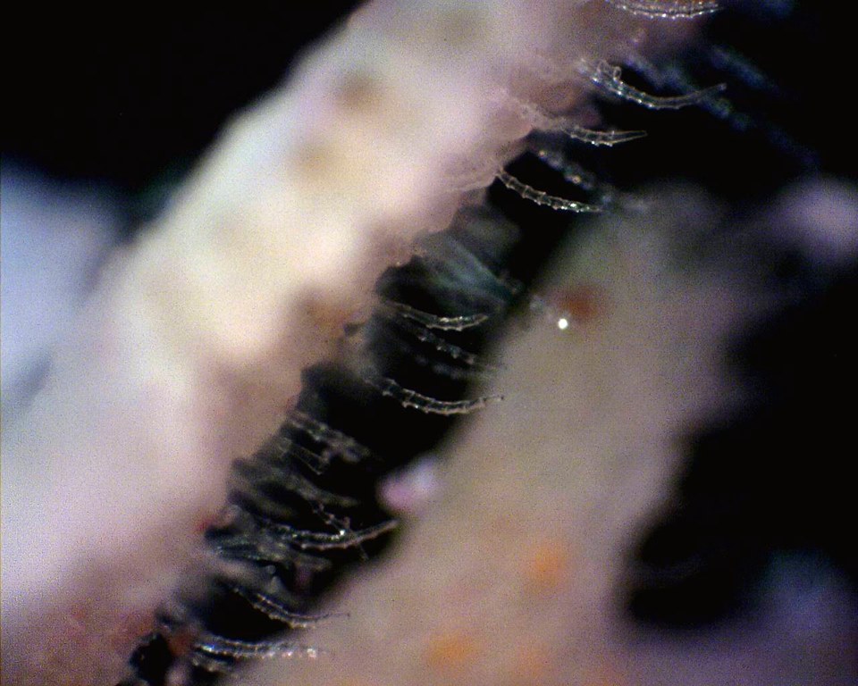
| Figure 2. R. graeffei, spicules. Photo: Bridget Bradshaw, Heron Island, 2013 |
The branches, or trabeculae, are slender with alternating series of 4 to 8 autozooids, each autozooid elongate and only 0.4 x 0.25 mm. The labial pore is squat and oval, the rim developing 2-3 spiked processes. The calcareous frontal shield facing outwards is smooth with raised sutures along the edges separating the individual zooecia. Avicularia can be found along the edges of the trabeculae and in the center of the frontal shield of most zooids (see SEM photos with full description here). Ovicells are dispersed infrequently throughout the colony and may be seen only in certain light, as they are clear and bubble-like attached on the periphery of the trabeculae (see REPRODUCTION).
The most common fenestrate bryozoan with which R. graeffei could be confused are Margaretta triplex and a species of Triphyllozoon, and all can occur on the same piece of coral rubble. Should there be no readily accessible SEM, the easiest way to distinguish R. graeffei from M. triplex is the difference in branching pattern, with R. graeffei branches fusing together as they grow to form a rippling network while M. triplex forms a bushier, more flexible colony with branches that don't grow together. Triphyllozoan sp. has a far more regular mesh appearance, with homologous fenestrulae (spaces between the branches) shaped like little diamonds.
|